Removal of chronic subdural hematomas in Ternopil

Removal of a chronic subdural hematoma is an operation to remove accumulated blood between the membranes of the brain, which over time slowly compresses the brain, causing impaired consciousness, weakness, or speech disorders. The surgeon makes a small hole in the skull (trepanation), through which the hematoma is removed and the cavity is irrigated, which instantly relieves the brain of pressure and restores normal well-being.

order a call
20+

years of experience in neurosurgery

3000+

operations performed

35+

internships in Europe

100%

modern techniques

Leontiev Oleksiy
INTERNSHIP SINCE 2002

LEONTIEV OLEKSIY

neurosurgeon of the highest category
candidate of medical sciences

head of the neurosurgical department
KNP "TOKPL" TOR

Value

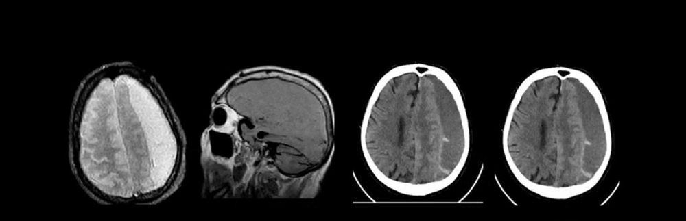
A subdural hematoma is a collection of blood that forms between the dura mater and the brain, the main cause of which is trauma. The main source of bleeding is damaged veins in the brain. A chronic hematoma is one that occurs more than 14 days after a traumatic brain injury.

The main feature that distinguishes an acute subdural hematoma from a chronic one is the presence of a connective tissue capsule that forms 2 weeks after TBI and separates the hematoma from the brain.

In most cases, patients do not seek medical help and can walk with this problem for several months or even years. Sometimes even minor blows to the skull can lead to these problems, and only at the height of clinical symptoms do patients seek medical help.

Symptoms

  • headache (which is most often of a “bursting” nature),
  • dizziness, nausea, vomiting,
  • speech impairment (the patient cannot say a word and/or does not understand the meaning of what is said),
  • weakness in the limbs on the opposite side of the stroke
  • epileptic seizures, mental disorders, memory impairment
  • meningeal syndrome.

hron chna subduralka kovalj 2

Diagnostic methods

ECHO-ES – in the absence of CT, MRI. The leading diagnostic methods are CT and MRI of the brain. Computed tomography is mainly performed in the acute period of injury because it better visualizes the accumulation of blood between the dural membrane and the brain.

In chronic subdural hematoma, MRI is preferred for diagnosis.

Indications for surgical treatment

  • hematoma volume (according to neuroimaging data) over 50 ml in volume.
  • dislocation of midbrain structures more than 5 mm
  • deformation of the basal nuclei,
  • hematoma volume (according to neuroimaging data) over 50 ml in volume.
  • gross compression of the lateral ventricles with the development of hydrocephalus, regardless of size and localization.

Surgical treatment of subdural hematomas in most cases, if there are no partitions in the hematoma structure, is carried out through 2 small holes in the bones of the skull, through which tubes for the inflow and outflow system are inserted, through which the remains of the hematoma are washed out during the day, and the drains are removed 2-3 days after the operation.

Frequently Asked Questions about removal of chronic subdural hematomas

All categories
How to get treatment according to the method removal of chronic subdural hematomas in Ternopil?

To get treatment, leave a request on the website via the “Make an appointment” button or call. The initial appointment is held at: Ternopil city, st. Trolleybusna, 14.

Patients’ procedure:

  1. Make an appointment: Fill out an application on the website or call the reception.
  2. Prepare documents: Bring your existing medical records and images (MRI/CT).
  3. Consultation: The neurosurgeon will conduct an examination, study the images, and determine the indications for the procedure.
Where is removal of chronic subdural hematomas performed in Ternopil?

Admission and procedure are carried out on the basis of a specialized department:

  • Institution: KNP “TOKPL” TOR (Neurosurgical Department).
  • Doctor: Leontiev Oleksiy Oleksandrovych – Head of the Department, Candidate of Medical Sciences.
  • Address: Ternopil, 14 Troleybusna Street.
  • Phone appointment: (073) 486 18 85
Leontiev Alexey — neurosurgeon
The article was reviewed by a medical specialist
Leontiev Alexey

neurosurgeon of the highest category

The information provided in this article, checked by a neurosurgeon of the highest category, Leontiev Alexey. It is of a general informational nature and does not replace the consultation of a specialist. For recommendations on diagnosis and treatment, a doctor's consultation is required.

ADDRESS
14, Trolleybusna St., Ternopil
OPENING HOURS
Mon-Fri: 8:30 AM – 5:30 PM
Precise diagnosis

Treatment planning is based on high-precision MRI, CT, and EEG images, which allows for a detailed assessment of the state of nervous structures even before the procedure begins.

Safety and Sterility Standards

Minimally invasive interventions are performed in the operating room in compliance with absolute sterility and under continuous X-ray or ultrasound control.

Popular destinations:

High precision Brain, spinal cord and nerve surgeries
Care and comfort Patient support at all stages of treatment
Modern techniques Minimally invasive technologies and international standards

Qualifications and certificates

Leave a Comment